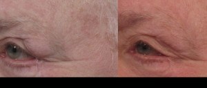
What is being treated?
A sagging or low-positioned tear gland can be elevated with a lacrimal gland suspension. This procedure will improve bulges seen at the outer ⅓ of the upper eyelid caused by a tear gland that has assumed a lower position than normal.
How is lacrimal gland suspension performed?
Incision
The skin is incised along the upper eyelid crease. This may be done at the same time as an upper blepharoplasty, with the same incision.
Structures are exposed
The surgeon dissects under the orbicularis muscle at the outer ⅓ of the upper eyelid, allowing for visualization of the lacrimal gland which consists of two lobes. The lacrimal gland fossa is also visualized, which is a recess just inside the bony orbital rim.
Gland suspension
The gland is stitched to the covering of the bone (periosteum) at the front of the lacrimal gland fossa. One or more stitches may be used.
Skin closure
The incision is closed with stitches.
Follow-up care
Stitches are removed at 5-10 days, during which swelling and bruising are expected.
Risks
Rare complications include bleeding, infection, dry eyes (due to interruption of tear flow or gland injury), asymmetry, and undercorrection.
Related procedures
Upper eyelid lift is often performed at the same time

